

3D pelvic ultrasound
3D pelvic ultrasound
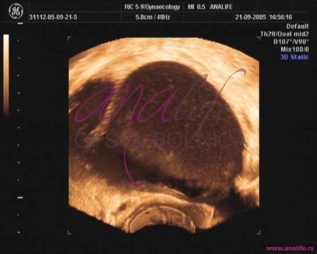
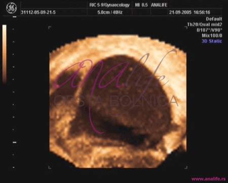
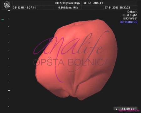
3D ultrasound enables a three-dimensional reconstruction of the internal genital organs.
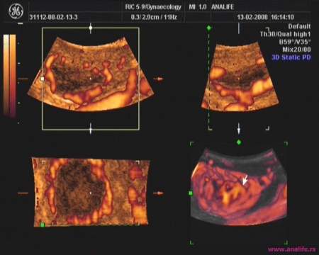
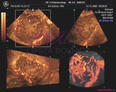
3D ultrasound enables a three-dimensional reconstruction of the internal genital organs (uterus, ovaries, and expanded fallopian tubes) and their blood vessels (3D power Doppler mode). This allows a spacious anatomical cross section analysis, which cannot be done with a classic 2D ultrasound. Most indications for combined 2D/3D US examination are diagnostic dilemmas after the standard 2D US.
Advantages of the 3D US over 2D US:
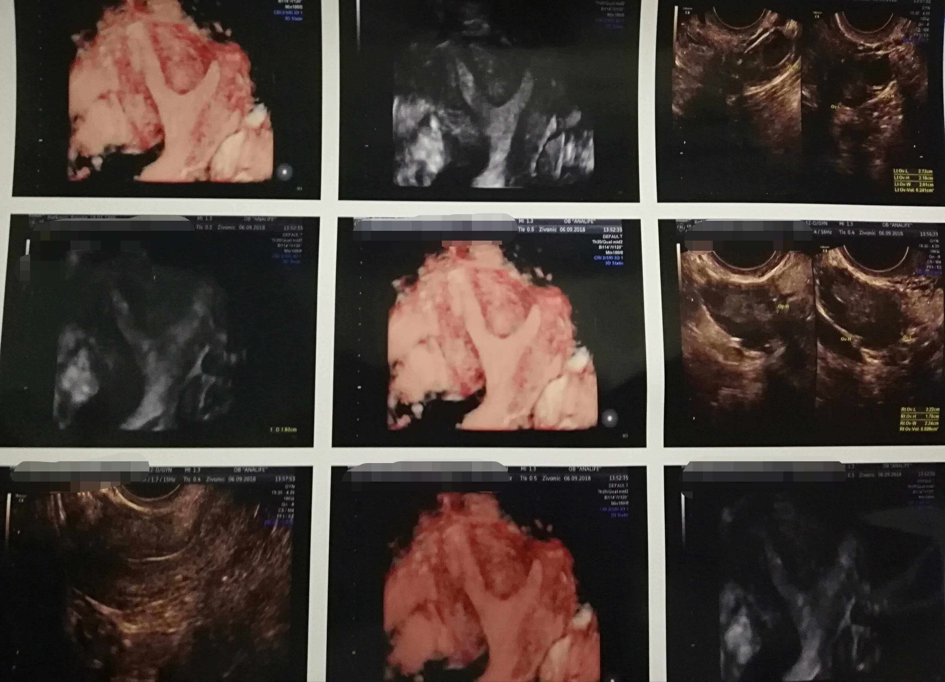
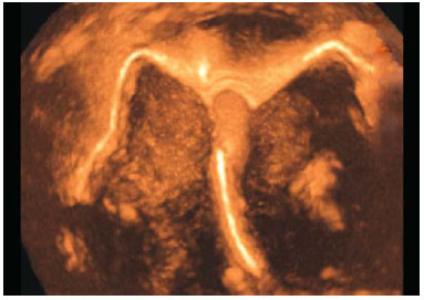
- Detection of congenital uterine anomalies (unicornuate, bicornuate, didelphic, septate, etc);
- Endometrium disorders;
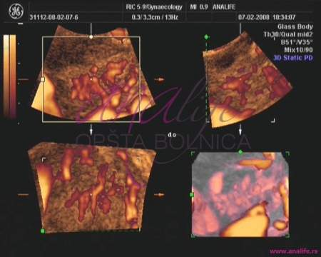
- The complete silhouette of the uterus lining is visible, which used to be possible only with hysterosalpingogram (HSG);
- Easier detection of polyps or myomas that are in the uterine cavity.
3D US is often useful addition to the standard US examination in the following:
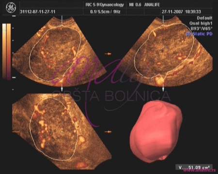
1.Myomas:
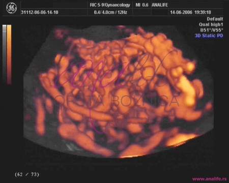
The size and location of the myomas can be precisely determined, as well as their blood supply and the tendency to grow.
On the basis of the acquired data, the decision is made regarding the type of the required surgical procedure.
2.Pelvic tumors:
It is easier to differ simple cysts from the real cystic tumors.
In the case of a real tumor, 3D location analysis, the size and characteristics of the tumor blood vessels indicate a risk of malignity.
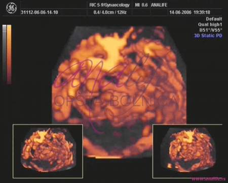
3.mage of the ovaries and their blood vessels in three dimensions:
Functionality of this important organ is assessed.
4.Examination of infertility causes:
Ovulation is easily viewed in the cycle in which the exam is done.
5.Polycystic ovaries:
Size, blood supply and functionality assessment.
6.Endometriosis:
Size, location and blood supply of the changes.
Based on the acquired data, the decision is made regarding the type of treatment (conservatory/ operative)
Note:
3D US is never done as an individual exam, but always combined with the standard 2D US or 2D Color Doppler. Before the US exam, the gynecologist has a professional obligation to perform the traditional pelvic exam. If you still do not want to have the pelvic exam done, in the conclusion of the US exam you will be advised to have the standard gynecological exam done.
Video gallery


Gallery


Monday-Friday from 09-20h
Saturday from 09-15h
Opšta Bolnica Analife
Skadarska 5
(Zemun), Beograd