

Gynecology
Gynecological (pelvic) exam
Gynecological examination starts by talking to the doctor and giving your medical history - talking about your health and reasons for coming, whether you have a gynecological problem, or you need a routine annual examination.
Basically, the exam includes three parts:
1.Visual exam:
-observation of the vulva with palpation of possibly detected changes.
2.Speculum exam:
- An instrument (speculum) is gently inserted into vagina to allow doctor to see the walls of vagina and the cervix.
3.Bimanual exam:
The gynecologist inserts two fingers of one hand into your vagina, and with the tips of the fingers of the other hand simultaneously presses down on the abdomen just above the pubic bone – in the region of the uterus and both ovaries. This way, the doctor can feel the changes in the uterus (enlarged uterus and even the reason for the enlargement, like pregnancy, myomas, etc.), and on the ovaries (inflammation, tumors…)
Ultrasound is a perfect addition to the gynecological exam. For this reason, pelvic US is considered a standard procedure when visiting a gynecologist. US is practically mandatory if the pelvic exam raises a doubt of present abnormality on the genital organs.
Virgin girls go to the rectal exam instead of the pelvic exam. In such cases, US needs to be done over the lower abdomen or rectally, which is recommended.
Colposcopy with PAPA smear needs to be done once a year, if the previous examination shows normal results.
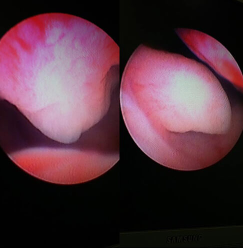
COLPOSCOPY with PAPA smear
- Colposcopy is a type of test where the uterus and vagina are inspected under a special microscope (colposcope). This enlarged image enables your doctor to analyze the surface of your cervix and vagina walls much clearer, and detect some suspicious changes. Just before the colposcopy, PAPA test is mandatory (these two methods are perfectly complementary).
Depending on the looks and scope of the detected changes, and the results of the PAPA test, the doctor will assess whether a biopsy is needed, to send a part of the tissue on the HP analysis.
Colposcopy with PAPA smear needs to be done once a year, if the previous examination shows normal results. This exam should be done for the first time not later than three years after the first sexual intercourse, and not later than the age of 21.
In modern gynecology, the following statement is generally confirmed and accepted:
Routine annual colsposcopic exam with PAPA test enables detection of premalignant and early stage malignant changes on the cervix, in the stage when they are completely curable with a smaller surgical procedure. In other words, this control method practically eliminates possibility of advanced forms of cervical cancer.
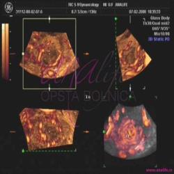
3D pelvic ultrasound
3D ultrasound enables a three-dimensional reconstruction of the internal genital organs.
3D ultrasound enables a three-dimensional reconstruction of the internal genital organs (uterus, ovaries, and expanded fallopian tubes) and their blood vessels (3D power Doppler mode). This allows a spacious anatomical cross section analysis, which cannot be done with a classic 2D ultrasound. Most indications for combined 2D/3D US examination are diagnostic dilemmas after the standard 2D US.
Advantages of the 3D US over 2D US:
- Detection of congenital uterine anomalies (unicornuate, bicornuate, didelphic, septate, etc);
- Endometrium disorders;
- The complete silhouette of the uterus lining is visible, which used to be possible only with hysterosalpingogram (HSG);
- Easier detection of polyps or myomas that are in the uterine cavity.
3D US is often useful addition to the standard US examination in the following:
1.Myomas:
The size and location of the myomas can be precisely determined, as well as their blood supply and the tendency to grow.
On the basis of the acquired data, the decision is made regarding the type of the required surgical procedure.
2.Pelvic tumors:
It is easier to differ simple cysts from the real cystic tumors.
In the case of a real tumor, 3D location analysis, the size and characteristics of the tumor blood vessels indicate a risk of malignity.
3.Image of the ovaries and their blood vessels in three dimensions:
Functionality of this important organ is assessed.
4.Examination of infertility causes:
Ovulation is easily viewed in the cycle in which the exam is done.
5.Polycystic ovaries:
Size, blood supply and functionality assessment.
6.Endometriosis:
Size, location and blood supply of the changes.
Based on the acquired data, the decision is made regarding the type of treatment (conservatory/ operative)
Note:
3D US is never done as an individual exam, but always combined with the standard 2D US or 2D Color Doppler. Before the US exam, the gynecologist has a professional obligation to perform the traditional pelvic exam. If you still do not want to have the pelvic exam done, in the conclusion of the US exam you will be advised to have the standard gynecological exam done.
Video gallery


Gallery

Monday-Friday from 09-20h
Saturday from 09-15h
Opšta Bolnica Analife
Skadarska 5
(Zemun), Beograd